
2. To develop an understanding of the relationships between cell components and clinical features of disease
3. To explain the bacterial growth curve
4. To familiarize you with immune reactions induced by the bacterial cell
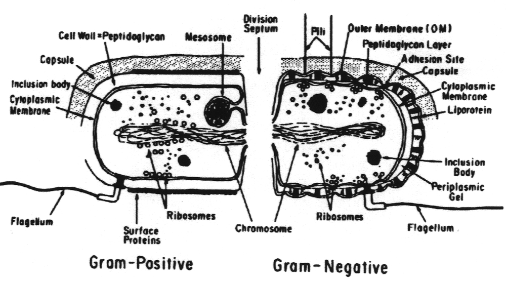
It is important to note the differences between the human (eukaryotic) cell and the bacterial (prokaryotic) cell because many of these differences account for disease pathogenesis and it has also been possible to exploit these differences in developing a chemotherapy regimen. In contrast to the human cell, the bacterial cell:
1.
May have a capsule. Not all bacterial cells have a capsule but when it
is present it is a major virulence factor. The capsule
includes the K-antigen.
2.
May have anouter membrane
which
is the outer surface of the cell or, in the case of encapsulated strains,
lies just
underneath the capsule. This has a trilaminar appearance. It contains lipopolysaccharides
(LPS). These are known
as endotoxins. They are also the somatic or O-antigen and are used in serological
typing of species. These occur only in
Gram-negative bacteria.
3.
May have a periplasmic
space which lies between the outer
membrane and the plasma membrane. This is filled with the
periplasmic gel which contains various enzymes. Again, this occurs only
in the Gram-negative bacteria.
4.
Has a rigid cell wall made of peptidoglycan (except for the mycoplasma).
This cell wall is thick in Gram-positive bacteria and
thin in Gram-negative bacteria. It is the thickness of the peptidoglycan
that accounts for the ability/lack of ability to retain the
crystal violet used in the Gram stain.
5.
Has a cytoplasmic
membrane lacking sterols (except for
the mycoplasma). Up to 90% of the ribosomes are attached to
this membrane. It also contains:
b. The membrane permeability (transport) systems.
c. Various polymer-synthesizing systems.
d. An ATPase.
7.
May have a flagellum which
arises from the plasma membrane and protrudes through the cell wall. This
is the source of the H
antigen which is used in serologic diagnosis. It is also the motility
organ and possibly an organ for attachment to a human
cell. It is considered a virulence factor.
8.
Has hairlike microfibrils, termed fimbriae
or
pili,
which originate in the plasma membrane and protrude through the cell
wall. They are straighter, thinner and shorter than flagella. The pili
contain chemical compounds called adhesins
which
allow the cell to bind to specific receptors on various human tissues.
This binding gives rise to organ specificity of some
bacterial strains. Fimbriae/pili are major virulence factors.
9.
Has ribosomes attached to the plasma membrane and also free in the cytoplasm
which have a mass of 70S (the human
ribosome has a mass of 80S). The protein and RNA species in the bacterial
ribosome differ from
those in the human ribosome.
10. May have an endospore within the cytoplasm. This is a body that allows the organism to resist adverse conditions.
11. Has a nucleus lacking a nuclear membrane.
12.
May have a circular plasmid.
This is a small (relative to the chromosome) piece of DNA that often codes
for virulence
factors.
13. Has a haploid (single) chromosome.
There are many common themes in bacterial pathogenicity related to cell structure of the species. These are based on the presentation to the human body of the bacteria, its parts and its metabolites. When an organism, or more commonly a number of organisms of the same species, enters the human body and encounters no host defenses, it will exhibit a growth curve like the one depicted below for a closed system.
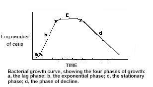
In the lag phase there is an increase in cell size at a time when little or no cell division is occurring. During this phase, there is a marked increase in macromolecular components (many of which are toxic to the human cell), metabolic activity and susceptibility to physical and chemical agents. The lag phase is a period of adjustment necessary for the replenishment of the cell's pool of metabolites to a level commensurate with maximum cell synthesis.
In the exponential orlogarithmic phase, the cells are in a state of balanced growth. During this state, the mass and the volume of the cell increase by the same factor in such a manner that the average composition of the cells and the relative concentrations of the metabolites remain constant. During this period of balanced growth, the rate of increase can be expressed by a natural exponential function.
The accumulation of waste products, exhaustion of nutrients, change in pH, induction of host immune mechanisms and other obscure factors exert a deleterious effect on the culture, resulting in a decreased growth rate. During the stationary phase, the viable cell count remains constant. The formation of new organisms equals the death of organisms in the system.
As the amount of the factors detrimental to the bacteria within the body increase, more bacteria are killed than are formed. During the phase of decline there is a negative exponential phase which results in a decrease in the numbers of bacteria within the system.
During all phases of the bacterial growth cycle, the host is exposed to the components of the bacterial cell. This exposure results in the induction of pathology as well as of immune mechanisms. The outcome is either life or death of the human, depending on the relative rates of induction of these phenomena.
The chemical nature of the capsule is important in the functions the capsule plays in the infection process. The capsules of bacteria are chemically diverse but the majority of them are polysaccharide in nature. These polymers are composed of repeating oligosaccharide units of two to four monosaccharides. Some may contain acetic acid, pyruvic acid and/or the methyl esters of hexoses. At least two species of pathogenic bacteria produce protein capsules; Bacillus anthracis produces a capsule of pure D-glutamic acid and Yersinia pestis produces a capsule of mixed amino acids. Capsules may be weakly antigenic to strongly antigenic, depending on their chemical complexity. Capsules may be covalently linked to the underlying cell wall or just loosely bound to it. Not all bacteria form capsules but in those that do the capsule is the interface between the bacterial cell and the external environment. As such it may serve a diversity of functions in disease including:
2.
Prevention of neutrophil killing of engulfed bacteria - lysosome contents
do not have direct access to the interior of the
bacterial cell and thus cannot kill the cell.
3. Prevention of complement-mediated bacterial cell lysis.
4.
Prevention of polymorphonuclear leukocyte migration to the site of infection
- Bacteroides fragilis produces a
polysaccharide capsule high in succinic acid. Succinic acid is released
from the capsule and paralyzes
the pmn leukocyte.
5.
Toxicity to the host cell - this takes many forms depending on the chemical
nature of the capsule. One example is the
capsule of B. fragilis which induces abscess formation.
6. Adhesion to the host cell.
7. Protection of anaerobes from oxygen toxicity.
8.
Determination of colonial type - bacteria with capsules form smooth (S)
colonies while those without capsules form rough
(R) colonies. A given species may undergo a phenomenon called S-R
variation whereby the cell loses the ability
to
form a capsule. Some capsules are very large and absorb water; bacteria
with this type of capsule (e.g., Klebsiella
pneumoniae) form mucoid (M) colonies.
9. Enhancement of the pathogenicity of other species in a mixed infection.
10. Receptors for bacteriophage.
11.
Induction of antibody synthesis - this is the basis for:
b.
Vaccine production. A polyvalent (23 serotypes) polysaccharide vaccine
of Streptococcus pneumoniae capsule is
available for high risk patients. There is also a polyvalent (4 serotypes)
vaccine of Neisseria meningitidis capsule
available. A monovalent vaccine made up of capsular material from Haemophilus
influenzae is also
available.
c. Quellung reaction
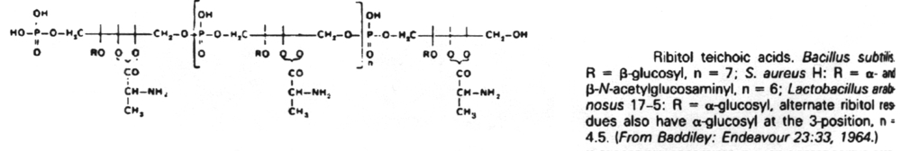
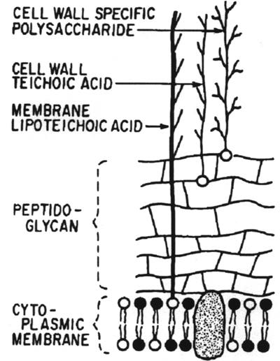
The capsule of bacteria may be penetrated by structures arising from the cell wall or plasma membrane such as cell wall specific polysaccharide, cell wall teichoic acid, plasma membrane lipoteichoic acid, flagella and pili.
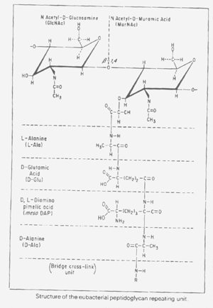
The glycan component is constituted of the two amino sugars, glucosamine and muramic acid. They occur as alternate ß-1, 4-linked N-acetyl-D-glucosamine and N-acetyl D-muramic acid residues. The glycan and peptide units are linked through the lactic acid carboxyl group of N-acetylmuramic acid to the amino terminus of a tetrapeptide. The glycotetrapeptides are cross-linked through the tetrapeptide units, forming a continuous 3-dimensional framework. While the tetrapeptide unit may vary with the species, the invariant feature of the tetrapeptide component is the presence of D-alanine, which is always the linkage unit between peptidoglycan chains.
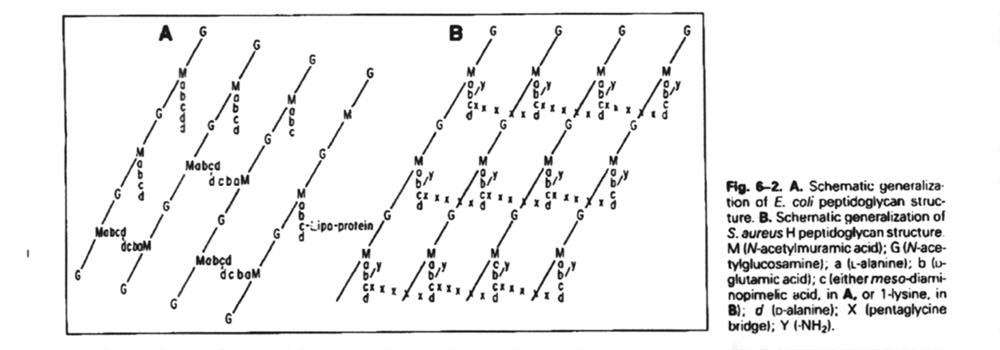
Thus, the cell wall can be several layers thick, each layer being a sheet of linked peptidoglycan units. The Gram-positive bacterial cell wall is distinguished by having multiple layers of peptidoglycan sheets and is thus up to ten times the thickness of a Gram-negative bacterial cell wall.
Attached to the rigid peptidoglycan framework of the cell wall are various polysaccharides which are covalently linked to the peptidoglycan. These fall into two groups:
C.
In some cases the cell wall of Gram-positive bacteria may contain proteins
of special significance. Examples of these
are:
A
composite of the cell wall of Gram-positive bacteria is diagrammed below.
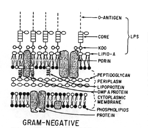
Between the cytoplasmic membrane and the outer membrane is the periplasmic space containing a gel-like periplasm in which resides the cell wall peptidoglycan as well as various enzymes.
In addition to phospholipids, the outer membrane contains unique Gram-negative lipopolysaccharides (LPS) and various proteins (porons) and lipoproteins. Each of these types of compounds is antigenic and is used to speciate and subspeciate organisms serologically. Of these compounds the LPS is the most important.
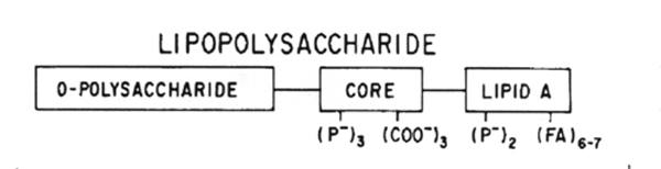
LPS is an amphiphile composed of three regions: O-polysaccharide (the O- or somatic-antigen), the core polysaccharide and lipid A. Lipid A is anchored in the outer membrane. LPS is also known as endotoxin.
The peptidoglycan of the Gram-negative cell is chemically similar to but not identical with the peptidoglycan of the Gram-positive cell. The major difference between the two cell types is in the thickness of the peptidoglycan rather than the chemical makeup.
When the bacterial cell wall is placed in the environment of the human body as part of a viable microorganism, there is a diversity of functions/effects that can be noted. Some of these are specific for Gram-negative organisms (due to the relative complexity of their cell walls) and some are general. The functions/effects of the cell wall include:
2.
Enhancement of the immune response to various cell metabolites by muramyldipeptide
(N-acetylmuramyl-L-alanyl-D-isoglutamine), i.e., it is an adjuvant
3. Induction of fever by muramyldipeptide (i.e., its a pyrogen)
4. Induction of sleep by muramyldipeptide (i.e., its a somnogen)
5.
Competition of muramyldipeptide with serotonin (5-hydroxytryptamine) for
receptors on macrophages. Serotonin, when
bound to the macrophage, enhances the chemotactic response of the macrophage.
Thus, muramyldipeptide blocks
this response in the inflammatory reaction.
6.
Induction of inflammatory arthritic joint disease by peptidoglycan-linked
polysaccharides (e.g., the polysaccharide of
group A streptococci linked to peptidoglycan)
7. Induction of granulomatous liver disease by peptidoglycan-linked polysaccharides
8. Stimulation of hemopoietic stem cells by peptidoglycan-linked polysaccharides
9.
Induction of chronic inflammatory bowel disease (i.e. Crohn's disease)
by peptidoglycan-linked polysaccharides,
especially those of Mycobacterium paratuberculosis.
10.
Induction of the immune response by the teichoic acids of Gram-positive
bacteria. This response is used in the
serological identification of Gram-positive bacteria.
11.
Induction of the immune response by the O-polysaccharide (somatic antigen)
portion of the lipopolysaccharide of the
outer membrane of Gram-negative bacteria. This response is used in the
serological identification of the Gram-negative
bacteria.
12.
Endotoxin (LPS) induction of:
B. Shwartzman
reaction - hemorrhagic necrosis at
the site of infection following exposure of another part of the
body to a relatively small amount of Lipid A. This is due to the clearing
of fibrin polymers at the inflammation
site.
C. Disseminated
intravascular coagulation - this can
lead to lethal shock. For this reason, it is especially
important in patients (e.g., with carcinoma) who suffer chronic disseminated
intravascular coagulation (defined
as a 10-20% decrease in circulating platelets and clotting factors).
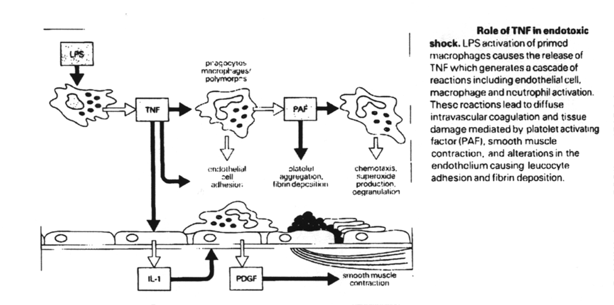
D.
Macrophage production of tumor
necrosis factor which
results in various effects including:
(2)
Adherence of polymorphonuclear leukocytes to the vascular endothelium,
causing them to degranulate
and form reactive oxygen intermediates such as superoxide anion and hydrogen
peroxide. This promotes
tissue necrosis and circulatory collapse.
F. Stimulation of bone marrow cell proliferation.
G. Nonspecific enhancement of immune responses (i.e., action as adjuvants).
H. Enhancement of radiation resistance
I. Clotting of horseshoe crab amebocyte lysates (Limulus lysate reaction).
J. Engender hypersensitivity reactions
13.
Functioning of the outer membrane of the Gram-negative cell wall as:
B. A molecular sieve for small water-soluble molecules.
C. An absorption site for bacteriophage
D. An absorption site for cellular conjugation
E. A reservoir for proteases, other enzymes and toxins
2.
Type I and type II pili promote adhesion to human cells with these results:
b.
Binding of bacterial cells to epithelial adhesion receptors which results
in interactions which may kill the human
cell. For example, Neisseria gonorrhoeae is avirulent if it lacks
pili.
2. All bacteria are classified as Gram-positive (retain the gram stain) or Gram-negative (do not retain the gram stain).
3.
Structural features of bacteria that are not seen in the human cell, or
differ from those in the human cell, include a capsule, an
outer membrane, a periplasmic space, a rigid cell wall, a cytoplasmic membrane
lacking sterols, the mesosome, flagellum,
fibrae (pili), 70S ribosomes, endospore, lack of a nuclear membrane, plasmids
and a haploid chromosome.
4.
The major antigens of the bacterial cell are the capsule (K-antigen), the
lipopolysaccharide (O-antigen) and the flagellum
(H-antigen).
5.
The growth cycle of a culture of bacteria is divided into four phases:
lag phase, exponential phase, stationary phase, decline
phase.
6.
The capsule of bacteria is most commonly polysaccharide in nature but proteinaceous
in at least two species, Bacillus
anthracis and Yersinea pestis.
7.
The capsule is a major virulence factor that allows bacteria to evade phagocytosis,
avoid the killing effects of lysosomal
enzymes, avoid complement-mediated cell lysis, paralyze leukocytes, induce
pathology in the host tissue, adhere to the host
cell, protect anaerobic cells from oxygen toxicity, produce a unique colony
type, enhance its pathogenicity, adsorb
bacteriophage and induce antibody synthesis.
8.
Bacteria with capsules from smooth (S) colones; those without a capsule
from rough (R) colonies; those with hydrophilic
capsules from mucoid (M) colonies.
9. Serologically, the capsule is important in diagnosis, vaccine production and as the basis for the Quellung reaction.
10.
The cell wall of bacteria is made up sheets of cross-linked repeating units
of peptidoglycan. In Gram-positive cells this is
relatively thick as compared to Gram-negative cells.
11.
Linked to the cell wall of bacteria are teichoic acids, cell wall specific
polysaccharides and, in some cases, proteins of special
significance.
12.
Gram-negative bacterial cells contain lipopolysaccharide (LPS) in their
outer membrane. This is the source of the O-antigen
and endotoxin.
13.
The functions/effects of the cell wall include maintenance of the morphology
or the bacterial cell, action as an adjuvant,
induction of fever, induction of sleep, competition with serotonin for
receptors on macrophages, induction of inflammation,
induction of liver granuloma, stimulation of hemopoietic stem cells, induction
of bowel inflammation, induction of antibody
synthesis.
14.
Endotoxin induces fever, hemorrhagic necrosis (Shwartzman reaction), disseminated
intravascular coagulation, production of
tumor necrosis factor, activation of the alternate complement pathway,
stimulation of bone marrow cell proliferation,
enhancement of the immune and the Limulus lysate reaction.
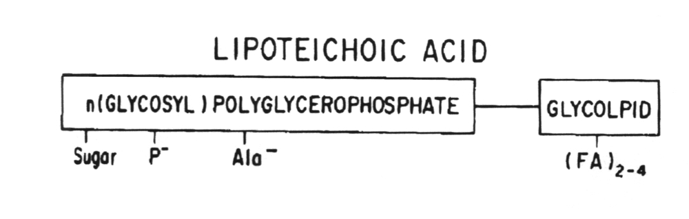
15. The lipoteichoic acid of Gram-positive bacteria acts similar to the endotoxin of Gram-negative bacteria.
16. Pili contain adhesins which allow the bacterial cell to bind to human cells.
17. Flagella are organs of locomotion that are used in serotyping strains of bacteria.