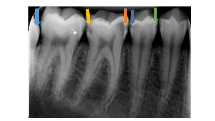
How would you stage these lesions? Light blue arrow, yellow arrow, orange arrow, dark blue arrow, and green arrow.
The ADA's 2015 Caries Classification System (CCS) for Clinical Practice
Dental caries are a continuum of net mineral loss. From a sound surface containing little to no mineral loss (Sound) to severe cavitation lesions containing a lot of mineral loss (Advanced).
All teeth and tooth surfaces should be visually inspected for lesions and can be scored using the table below (CCS). Not all carious lesions are active. Once a lesion has been identified and determined to be an Initial, Moderate or Advanced lesion then each lesion should evaluated using Table 1 to determine if the lesion is active or inactive. Treatment decisions can then made based on the CCS scoring and if the lesion is active or inactive. Definitions of tooth surface site definitions are below in Table 2.
2015 ADA Dental Caries Classification System (CCS)* |
||||
Sound |
Initial |
Moderate |
Advanced |
|
Clinical Presentation |
No clinically detectable lesions, Dental hard tissue appears normal in color, translucency and gloss. |
Earliest clinically detectable lesion compatible with mild demineralization. Lesion limited to enamel or to shallow demineralization of cementum/dentin. Mildest forms are detectable only after drying. Established and active lesions may be white or brown and enamel has lost its gloss. |
Visible signs of enamel breakdown or signs the dentin is moderately demineralized. |
Enamel is fully cavitated and dentin is exposed. Dentin lesion is deeply/severely demineralized. |
| Other Labels | No surface change or adequately restored |
Visually noncavitated |
Established early cavitated, shallow cavitation, or microcavitation |
Spread/disseminated, late cavitated, deep cavitation |
| Infected Dentin | None |
Visually noncavitated |
Possible |
Present |
| Appearance of Occlusal sufaces (Pit and Fissure or Smooth surfaces including Cervical and Root) | ICDAS 0** |
ICDAS 1 or ICDAS 2 |
ICDAS 3 or ICDAS 4 |
ICDAS 5 or ICDAS 6 |
| Radiographic Presentation of the Approximal Surface | E0*** or R0# No radiolucency |
E1 or RA1 OR E2 or RA2 OR D1 or RA3 Radiolucency may extend to the dentinoenamel junction or outer one-third of the dentin. Note: radiographs are not reliable for mild occlusal lesions. |
D2 or RB4 Radiolucency extends into the middle one-third of the dentin. |
D3 or RC5 Radiolucency extends into the inner one-third of the dentin. |
** For ICDAS system click here. ***E0-E2, D1-D3 notation system; E0 (no lesion), E1 (lesion within the outer half of enamel), E2 (inner half of enamel), D1 (outer third of dentin), D2 (middle third of dentin), and D3 (inner third of dentin); click here for article (Anusavice K. Present and future approaches for the control of caries. J Dent Educ. 2005;69(5):538-854.) #R0,RA-1-RA-3, RB4, and RC5-RC6 is the ICCMS radiographic scoring system (RC-6 = into pulp; https://www.icdas.org/uploads/ICCMS-Guide_Full_Guide_US.pdf . Accessed 9/22/16) |
||||
The American Dental Association Caries Classification System for Clinical Practice
Douglas A. Young, DDS, EdD, MBA, MS, Brian B. Nový, DDS, Gregory G. Zeller, DDS, MS, Robert Hale, DDS, Thomas C. Hart, DDS, PhD, Edmond L. Truelove, DDS, MSD Kim R. Ekstrand, DDS, PhD, John D.B. Featherstone, MSc, PhD, Margherita Fontana, DDS, PhD, Amid Ismail, BDS, MPH, DrPH, MBA, John Kuehne, DDS, MS, Chris Longbottom, BDS, PhD, Nigel Pitts, BDS, PhD, David C. Sarrett, DMD, MS, Tim Wright, DDS, MS, Anita M. Mark, Eugenio Beltran-Aguilar, DMD, DrPH, DABDPH Douglas A. Young, DDS, EdD, MBA, MS, Brian B. Nový, DDS, Gregory G. Zeller, DDS, MS, Robert Hale, DDS, Thomas C. Hart, DDS, PhD, Edmond L. Truelove, DDS, MSD Kim R. Ekstrand, DDS, PhD, John D.B. Featherstone, MSc, PhD, Margherita Fontana, DDS, PhD, Amid Ismail, BDS, MPH, DrPH, MBA, John Kuehne, DDS, MS, Chris Longbottom, BDS, PhD, Nigel Pitts, BDS, PhD, David C. Sarrett, DMD, MS, Tim Wright, DDS, MS, Anita M. Mark, Eugenio Beltran-Aguilar, DMD, DrPH, DABDPH. The Journal of the American Dental Association. Volume 146, Issue 2, Pages 79-86 (February 2015) DOI: 10.1016/j.adaj.2014.11.018
|
How would you stage these lesions? Light blue arrow, yellow arrow, orange arrow, dark blue arrow, and green arrow. |
© 2016 Neal R. Chamberlain,Ph.D. All rights reserved.
Site Last Revised 9/23/16
Neal Chamberlain, PhD. A. T. Still University of Health Sciences/Kirksville College of Osteopathic Medicine.
Take Care and Think Microbiologically!
Disclaimer:
None of the contributors to this web site assume liability for any use or misuse of the information provided here. This information does not replace dental/medical consultation, but is intended for EDUCATIONAL purposes only. Any mention of commercial products is for inclusive purposes and is not an endorsement. Every effort has been made to provide accurate information about the products mentioned in this web site, but consult product labels for the most up-to-date information available. This web site does not constitute a medical diagnosis; seek advice from a dentist/physician (if you are not one yourself) before making a diagnosis and/or undertaking any of the treatments suggested here.