Figure 1 |

|
|
In the emergency room, he had two episodes of vomiting and one episode
of diarrhea. His temperature was 38.9oC, pulse 160 beats per
min, and respiratory rate 36/min, and he was noted to be dehydrated. His
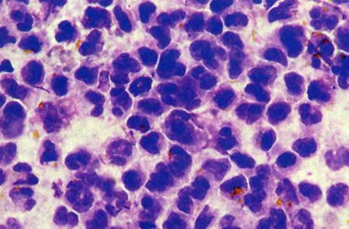
stool was mucoid and contained bloody streaks. A methylene blue stain of
his feces is shown below in figure 1. Other laboratory
studies included a cerebrospinal fluid examination, done because of his
lethargy, which was within normal limits; a peripheral white blood cell
count of 13,200/microliter with 85% neutrophils; a negative blood culture;
and a negative stool examination for ova and parasites. Figure
2 shows a MacConkey agar plate culture of the organism recovered from
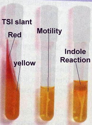
the feces of the patient. Figure 3 shows the biochemical
reactions obtained in a triple sugar iron (TSI) slant, a urea-motility-indole
(UMI) tube (look for motility), and a UMI with added Kovac's reagent (to
detect indole production; red positive; yellow negative).
|
Figure 1 |
|
|
To obtain the answers to these questions click this link: answers.
1. Why were the white blood cells present in his stool?
2. Based on the laboratory results seen in figures 1, 2, and 3, what organism is likely causing this illness (I only need the genus)?
3. What was the significance of his being in a group daycare?
4. What special characteristics of this organism lead to its spread?
Take Care and Think Microbiologically!
Neal R. Chamberlain, Ph.D.
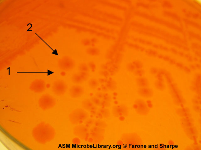
The small colonies (arrow number 1) of the strain of Shigella sonnei are more virulent whereas, the bigger colonies (arrow number 2) are less virulent. The O polysaccharide covalently attached to the LPS in the outer membrane causes the colonies to be small and makes the organisms resistant to complement-mediated lysis and phagocytosis.
Answers 1= The organism that is causing this person's current problem induces a strong inflammatory response in the intestine as a result of its infection of the cells lining the intestine.
2= Shigella MacConkey agar only grows gram negative rod shaped bacteria and the organism growing here is not a lactose fermentor (colonies would be pink to light purple if lactose fermentor). This result points toward Salmonella or Shigella as the etiological agent. In figure 3 the medium in the middle demonstrates the organism is not motile. The medium on the left (TSI agar) demonstrates that the organism is not a lactose fermentor (red slant; yellow butt) nor does it produce hydrogen sulfide gas (H2S gas caused the medium to turn black). Salmonella are usually motile and also produce H2S. The organism is unlikely to be E. coli because E. coli is a lactose fermentor and is indole positive. Therefore the most likely genus is Shigella which is nonmotile, indole negative, and H2S negative.
3= Very few organisms if ingested are needed to cause an infection with this bacterium (around 200). Since these children are not yet able to use the bathroom to defecate someone must change their diapers. If a person does not or cannot wash their hands after changing a child's diaper and then prepares a drink or food for another child or touchs another child in such a way that the organisms are consumed this other child can be infected. Shigella infections and the spread of these infections is more common in institutional settings like daycares, nursing homes, and prisons.
4= Its ability to cause infection when small numbers of the organism are ingested (around 200 organisms).
This site is maintained by Neal R. Chamberlain, Ph.D., and was last revised 1/13/09.
Comments can be emailed to: nchamberlain@atsu.edu
Copyright© 2002-2009, A.T. Still University/ Kirksville College of Osteopathic Medicine, Kirksville MO.