
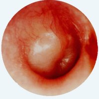
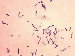
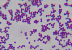
Gina brings Geraldine in with a high fever. Geraldine's vital signs; temperature- 104 degree C, heart rate- 140, respiratory rate- 23, bp- 120/80. When asked where it hurts Geraldine points to her left ear. Examination reveals left-sided cervical lymphadenopathy. Using an otoscope you see the image in figure 1 below. Geraldine is not happy when you put air in her ear to test for tympanic membrane mobility (did not move). She pushes the pneumatic otoscope out of her ear and starts crying. Her crying intensifies until suddenly she says she feels something in her left ear and it does not hurt as much. You note a bloody discharge draining from her ear when you position her head to allow drainage of the left ear. Before she can run away again you obtain a sample of the material coming out of her ear for culture. Otoscopic exam reveals tympanic membrane rupture. Growth on blood agar plates reveals three different colony types. Each colony type was gram stained (Figures 3, 4, and 5). Various biochemical tests were performed to identify the organisms (see table containing the figures).
Figure 1; Left Ear ; no tympanic membrane mobility; no cone of light |
Figure 2; Right Ear; good tympanic membrane mobility and cone of light |
 |
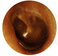 |
Figure 3; Aerobic growth on blood agar with no hemolysis |
Figure 4; aerobic growth on blood agar with alpha hemolysis, catalase negative |
Figure 5; aerobic growth on blood agar with no hemolysis, catalase negative, coagulase negative |
 |
 |
 |
A. Which of the images above is the most likely cause of Geraldine's current condition?
1. Figure 3- a gram positive rod; likely a diphtheroid (a skin dwelling Corynebacteria species)- notice the broken stick or Chinese letter morphology and the uneven staining.
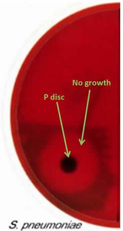
2. Figure 4- Three most common causes of otitis media are Streptococcus pneumoniae (gram positive coccus) , Haemophilus influenzae (gram negative rod) , and Moraxella catarrhalis (gram negative coccus); This organism is a gram positive coccus that is alpha hemolytic on blood agar plates. If a P disc sensitivity test had been done on blood agar you would have seen no growth around the P disc (see Figure 6 below).*
3. Figure 5- a gram positive coccus; catalase positive makes it likely to be a Staphylococcus; coagulase negative makes it likely to be Staphylococcus epidermidis
B. Match the Figure number with the most likely name for the organism in that figure.
Organisms; Streptococcus pneumoniae, Corynebacterium sp., Staphylococcus epidermidis
Figure # |
Organism |
3 |
Corynebacterium sp. |
4 |
Streptococcus pneumoniae |
5 |
Staphylococcus epidermidis |
Figure 6; no growth around P disc |
 |
© 1996-2013 Neal Chamberlain. All rights reserved.
Site Last Revised 11/12/13
Neal Chamberlain, PhD. A. T. Still University of Health Sciences/Kirksville College of Osteopathic Medicine.
Take Care and Think Microbiologically!