|
|
|
|
CHOLERA
ETIOLOGICAL AGENT:
Vibrio cholerae. This is a slightly curved gram-negative rod which has two major groups based on the O-antigen. These are identified by slide agglutination tests with specific antiserum. Classic epidemic cholera is caused by the 01 serotype; all other strains are designated the non-01 strains and they have the antigen designations 02-0139. These non-01 strains produce sporadic and milder forms of diarrhea.
OVERVIEW:
Cholera is endemic in India, West Bengal, Bangladesh
and Louisiana in the U.S. The organism is ingested with water or food (especially
shellfish and crabs) and causes an acute illness due to an enterotoxin
elaborated by V. cholerae that have colonized the small bowel. In
its most severe form, there is rapid loss of liquid and electrolytes from
the gastrointestinal tract, resulting in hypovolemic shock, metabolic acidosis
and, if untreated, death.
PATHOLOGY:
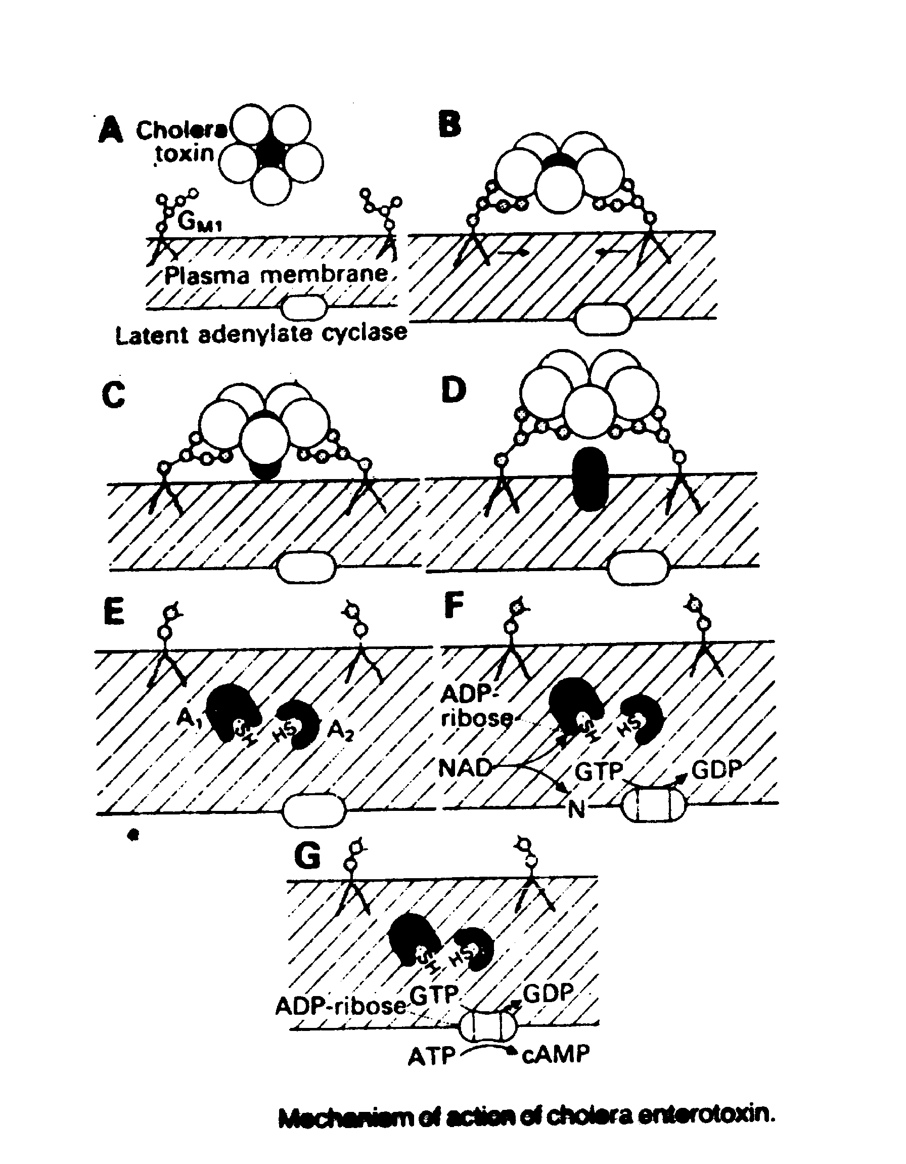
V. cholerae is very acid sensitive and the majority of ingested organisms are killed by stomach acidity; it takes ingestion of 108-1010 cells to cause disease. Those few organisms that survive stomach and upper intestinal acidity attach to the microvilli of the brush border of epithelial cells of the jejunum and ileum. There they multiply and liberate cholera enterotoxin, mucinase and endotoxin. They do not invade the mucosa. All signs, symptoms and metabolic derangements in cholera result from the rapid loss of liquid from the gut. The feces are nearly isotonic with plasma. As compared with plasma, the concentrations of sodium and chloride are slightly less, bicarbonate is twice as high, and potassium is 3-5 times higher. The increased electrolyte secretion is caused, in the absence of morphologic damage to the gut mucosa, by a protein enterotoxin coded for by a chromosomal gene. The enterotoxin has a molecular mass of 84,000 daltons and consists of a binding (B) moiety and an activating (A) moiety.
Five equal subunits with a molecular weight of 11500 each make up the B moiety.
On exposure to small bowel epithelial cells, each
B subunit rapidly binds to GM1 monosialoganglioside in the gut
cell wall. Following binding, the A moiety (composed of two unequal subunits)
migrates through the epithelial cell membrane. The A1 subunit
contains ADP-ribosyltranferase activity and catalyzes the transfer of ADP-ribose
from NAD to a guanosine triphosphate (GTP) - binding protein that regulates
adenylate cyclase activity. The ADP-ribosylation of GTP binding protein
inhibits the GTP turnoff reaction and causes a sustained increase in adenylate
cyclase activity. The resultant increased intracellular cyclic AMP acts
at 2 sites to cause net secretion of isotonic liquid within the small bowel
lumen. The increased cyclic AMP inhibits neutral sodium chloride absorption
across the brush border via the cotransport mechanism; it also stimulates
active chloride secretion into the gut lumen. There is no significant pathology.
CLINICAL SYMPTOMS:
The onset is characterized by abrupt, painless, watery
diarrhea. Several liters of liquid may be lost within a few hours, rapidly
leading to profound shock. Vomiting may ensue after diarrhea. The patient
is cyanotic and has sunken eyes and cheeks, a scaphoid abdomen, poor skin
turgor and thready or absent peripheral pulses. The voice is high pitched
or inaudible; the vital signs include tachycardia, tachypnea and low or
unobtainable blood pressure. The heart sounds are distant and often inaudible,
and bowel sounds are hypoactive.
DIAGNOSIS:
In endemic or epidemic areas, the working diagnosis
of cholera is made based on the clinical presentation, especially the presence
of "rice water" stools. Confirmative diagnosis is made by plating a stool
sample on TCBS (thiosulfate-citrate-bile salt-sucrose) agar, which is selective
for
Vibrio, and the adrenal cell assay.
TREATMENT:
Successful therapy requires only prompt replacement
of fluids and electrolytes. Ringer's solution is most commonly used. It
is given rapidly by IV injection - 50 to 100 ml per minute - until a strong
radial pulse is restored. Tetracycline reduces the severity and length
of disease. Chloramphenicol and furazolidone are slightly less effective.
PREVENTION:
A parenterally administered vaccine containing 109
killed vibrios per ml is available. Protection lasts for 3-6 months. It
is effective only against 01 serotypes.
ESCHERICHIA COLI INFECTION
ETIOLOGICAL AGENT: Enterotoxigenic E.
coli (ETEC) or
enteropathogenic E. coli (EPEC)
PATHOLOGY:
EPEC produces no demonstrable toxin but most strains are enteroadherant (EAEC) and cause alterations in the brush border of the small bowel epithelial cells. EPEC express rope-like bundles of filaments, termed bundle-forming pili, that create a network of fibers that bind together the individual organisms. The EPEC pili are homologous with toxin-coregulated pili of Vibrio Cholerae. The gene for these fibers is plasmid-borne. ETEC has multiple pathogenic mechanisms which include at least 2 distinct toxins; a heat-labile toxin (LT) and a heat stable toxin (ST). The ability to produce enterotoxin is mediated by a single plasmid. The E. coli LT is similar, but not identical to, the cholera toxin; it binds to the same site, has the same effect and serologically cross-reacts with cholera toxin. Like cholera toxin it will bind to cells outside the intestine.
The E. coli ST is quite different from the
LT because it exhibits a rapid onset of action, does not bind to gangliosides
of the mucosal cell membrane, and is of low molecular weight, that is,
less than 2000 daltons. It is not antigenic. The ST acts by stimulation
of guanylate cyclase with resultant cyclic GMP accumulation in mucosal
cells. The kinetics of ST action are strikingly different from LT action,
because it causes an almost immediate increase in the secretion of gut
fluid. The increase in intracellular guanylate cyclase causes chloride
secretion by gut mucosal epithelial cells in a manner similar to that seen
with the cholera enterotoxin, but the ST does NOT alter neutral sodium
chloride absorption by the brush border cotransport route. ST binding is
specific for small bowel mucosal cells and thus, unlike cholera enterotoxin
and E. coli LT, it affects only intestinal cells. Besides toxin
production, the major other violence factors that EPEC and ETEC produce
are colonization factors which allow them to adhere to mucosal cells. These
are various types of fimbriae or pili. These strains produce little or
no gross pathology.
SYMPTOMS:
Severe diarrheal disease caused by ETEC is generally characterized by the abrupt onset of watery diarrhea. In severe cases, the clinical picture is identical to that of cholera except that cramping abdominal pain is more commonly present with E. coli diarrheas and the duration is much less, seldom lasting more than 24 hours after initiation of fluid replacement therapy. The non-enterotoxin producing, noninvasive E. coli have thus far been incriminated only in relatively mild diarrheal disease in infants and small children.
DIAGNOSIS:
Diagnosis is made by isolating E. coli on MacConkey's agar and then:
1.
Inoculating them into a tissue culture of mouse adrenal cells or Chinese
hamster ovary
cells which respond morphologically to stimulation of their adenylate cyclase
systems by
the LT or
2. Performing an ELISA test on toxin bound to antibody or
3.
Using a DNA probe to detect the LT gene.
TREATMENT:
Intravenous or peroral replacement of the fluid and
electrolytes lost in feces. Peroral therapy is almost always adequate.
Tetracycline and trimethoprim - sulfamethoxazole are effective in shortening
the duration of symptoms but are not essential. Bismuth subsalicylate may
provide symptomatic relief (less severe abdominal cramps and less frequent
stools).
GIARDIASIS
NAME OF DISEASE: Giardiasis
ETIOLOGICAL AGENT:
Giardia lamblia, a flagellate with both a
trophic and cystic stage. The teardrop-shaped trophozoites have a smooth
dorsal surface with a concave ventral surface and a prominent anterior
adhesive disk. There are 4 pairs of flagella directed posteriorly. The
cysts are ellipsoidal and highly refractile. This is the most common intestinal
protozoan parasite of humans.
PATHOLOGY:
Ingested organisms colonize the duodenum and jejunum
where they injure the epithelium of the microvillus, which is manifested
as a functional derangement in the cell membrane and a loss of the glycocalyx
coat. Loss of the digestive enzymes and transport mechanisms for mono-saccharides
and amino acids leads to malabsorption and diarrhea. Pathologic changes
are mild in most cases, but shortening and thickening of the villi associated
with acute focal inflammatory changes in the mucosal epithelium may be
seen initially and are followed by chronic inflammatory infiltrates in
the lamina propria.
TREATMENT:
Quinacrine hydrochloride (Atabrine; 100 mg, PO, every
8 hours for 10 days) or metronidazole (Flagyl;250 mg, PO, every 8 hours
for 10 days).
ANTIMICROBIC-ASSOCIATED PSEUDOMEMBRANOUS COLITIS
Clostridium difficile is an anaerobic, spore-forming,
gram-positive bacillus that is normal flora in about 5% of humans. Its
numbers are held in check by other normal flora so that it does not normally
cause disease. It is resistant to almost all antibiotics and may grow to
huge numbers when antimicrobics inhibit bacterial competitors. In this
case it produces sufficient toxin of two types to cause a pseudomembranous
colitis. The pseudomembrane consists primarily of inflammatory cells plus
fibrin and is adherent to the denuded surface of the distal colon. The
intervening mucosa is marked by hemorrhagic inflammation. Withdrawal of
antibiotics clears the condition.
VIRAL GASTROENTERITIS
There are several viruses that may cause gastroenteritis. One of these, the rotavirus, is the most important cause of infantile gastroenteritis around the world. It causes 5 million deaths of preschool children annually. This is a member of the Family Reoviridae (naked, icosahedral viruses with segmented double-stranded RNA). Rotaviruses infect epithelial cells of the villi where they multiply in the cytoplasm and damage the sodium and glucose transport mechanisms, thus inducing diarrhea. Up to 1010 viruses/gm occur in the feces. Typical symptoms are diarrhea, fever, abdominal pain and vomiting. This has been termed "winter" diarrhea. Treatment is replenishment of fluids and electrolytes. Prevention is via the administration of three oral doses of a live attenuated quadrivalent vaccine at 2, 3 and 4 months of age.
The second most common cause of viral gastroenteritis
is the Norwalk Agent, a Calicivirus (naked, icosahedral virus with single-stranded
RNA). This virus causes "summer" diarrhea with typical gastroenteritis
symptoms.
|
|
|
|