|
|
|
|
DERMATOLOGIC INDICATIONS OF INFECTION
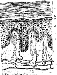
The integument or skin refers to the outer covering of the body which includes the stratum corneum, epidermis, dermis and the various structures integrated into this tissue. These structures consist of sweat glands, hair follicles, sebaceous glands, blood vessels, ducts and peripheral nerves. Specialized stratum corneum cells make up the nails and hair shafts. The integument has the ability to react to infection in a variety of ways which allows the physician knowledgeable in these reactions to diagnose both external and internal infectious diseases with greater accuracy and speed. In performing the dermatological examination the following data should be obtained:
I. History
A. Family history of dermatologic disorders
1. Pigment disorders (albinism, melanoma, vitiligo, freckles, etc.)
2. Neurocutaneous diseases (von Recklinghausen's
disease, tuberous sclerosis,
Sturge-Weber syndrome) all involve depigmentation and paralysis of localized
areas
of the body.
3. Vascular diseases (Rendu-Osler-Weber syndrome,
angiokeratomas,
Wiskott-Aldrich syndrome, Milroy's disease)-all involve hemorrhage into
the skin.
4. Diseases of the corium (cellulitis, peripheral neuritis, hidradenitis).
5. Diseases with prominent epidermal reactions
(ichthyosis, pityriasis eczema,
keratosis, psoriasis, allergy).
6. Metabolic diseases (porphyria, amyloidosis, lipoid proteinosis, gout).
7. Tumors (keloids, nevi, warts).
B. Patient history
1. General review
a. Occupation
b. Exposure to toxic agents
c. Nutrition
2. Past dermatologic history
3. Present illness
a. Chief complaint
b. Location of initial lesion
c. Evidence of progression
d. Distribution of lesions on the body
(1) Extremities only
(2) Trunk only
(3) Palms and soles
(4) Head and face
(5) Skin folds only
(6) Mucous membranes
e. Contact with infected person
II. Examination
A. Distribution of rash
B. Color of rash
C. Size of rash. Discrete or confluent? Shape?
D. Are lesions elevated, even with the plane of the skin or depressed?
E. Surface texture of rash
F. Vascular or extravascular reaction
G. Persistence of rash
H. Color of rash upon application of pressure
I. View of rash under Wood's lamp
J. Palpation of lesion (soft, doughy, hard, infiltrated, dry, moist, change in temperature)
K.
Type of lesion (see table 1)
| Table 1 Types of skin lesions | ||
| Flat lesions
(in the plane of the skin) |
Elevated lesions
(above the plane of the skin) |
Depressed
lesions (below the plane of the skin) |
| Macule
IInfarct Sclerosis Telangiectasis |
Vesicle and bulla
Pustule Abscess Cyst Papule Wheal plague Nodule Vegetation Keratosis Desquamation (scales) Exudate (crusts) Lichenification |
Atrophy§
Sclerosis§ Erosion Excoriation Scar Ulcer Sinus Gangrene§ |
| May also be below the plane of skin
May also be above the plane of skin May also be in or below the plane of skin § May also be in the plane of skin |
||
L. Morphologic component(s) of skin primarily affected (see Table 2)
| Table 2 Clinical classification of skin lesions and syn-dromes according to the component of the skin primarily involved |
| I. Epidermis (keratinocytes and metanocytes)
A. Keratinocytes 1. Scaling macules, papules, or plaques 2. Vesicles and bullae 3. Pustules 4. Exudative (impetiginized) lesions 5. Eczematous dermatitis 6. Erythroderma syndrome (exfoliative dermatitis) 7. Atrophy, diffuse or circumscribed B. Metanocytes 1. Hypomelanotic macules 2. Diffuse hypometanosis 3. Hyper melanotic (brown macules) 4. Diffuse brown hpermelanosis II. Dermis (connective tissue and blood vessels) A. Connective tissue component 1. Papules and nodules (with and without inflammation) 2. Ulcers 3. Sclerosis, diffuse or circumscribed 4. Edema 5. Atrophy, diffuse or circumscribed B. Blood vessels 1. Morbilliform and scarlatiniform eruptions 2. Unticanal syndromes 3. Erythema multiform syndrome 4. Purpura (with end without inflammation) 5. Infarcts 6. Telangiectasia III. Panniculus adiposis (connective tissue and blood vessels) A. Connective tissue component 1. Nodule, nonflammatory, usually non tender 2. Atrophy B. Blood Vessel 1. Nodules, inflammatory, usually tender and red a. Erythema nodusum syndrome |
| Pathologic changes after large areas of skin, and there are no ciscrate circumstance reasons. |
M. Sensation (pain, itchy, paresis)
N. Nikolski's sign
O. Are lesions all at the same stage of development?
P. Is edge of lesion well defined? Smooth or irregular?
Q. Schultz-Charlton Test
III. Laboratory Tests
A. Special procedures
1. Gram stain of material (crusts, scales, exudate) for bacteria.
2. KOH treatment of material for fungi
3. If vesicle, direct smear of base for multinucleated
giant cells (may indicate herpes
virus infection). This is the Tzanct test.
4. Biopsy for bacterial and mycologic culture.
5. Wood's lamp examination of urine for porphrins and hair and skin for fluorescence.
B. General procedures-hematologic, chemical, serologic, roentgenologic, urinalysis.
IV. Definition of terms
Abscess-a localized collection of pus caused by suppuration in a tissue or confined space.
Atrophy-wasting or disintegration of tissue.
Bulla-a
circumscribed, fluid containing, elevated lesion of the skin more than
0.5 cm in
diameter.
Cyst-a pathologic, epithelium-lined cavity usually containing fluid or semisolid material.
Desquamation-shedding of cells from the skin (scale formation).
Erosion-a wasting away of the skin, a kind of ulceration.
Excoriation-removal of an area of the skin.
Exudate-material
which has escaped from blood vessels and been deposited in tissues or on
tissue surfaces, usually as a result of inflammation.
Gangrene-death
of tissue in considerable mass, usually associated with loss of vascular
supply.
Infarct-a
localized area of ischemic necrosis produced by occlusion of the arterial
supply
or the venous drainage.
Keratosis-a horny growth of tissue.
Lichenification-thickening and hardening of the skin.
Macule-a
circumscribed area of change in normal skin color without elevation or
depression
relative to the surrounding skin.
Maculopapule-a papule developed on a macule
Nodule-a
palpable, solid , round or ellipsoidal lesion which lies deeper in the
skin than a
papule.
Papule-a
solid lesion generally considered to be less than 1 cm in diameter, which
is
elevated above the plane of the surrounding skin.
Plaque-an
elevated lesion that occupies a relatively large surface area (more than
1 cm in
diameter) and is frequently formed by the confluence of papules.
Pustule-a circumscribed, pus-containing lesion of the skin, up to 0.5 cm in diameter.
Scar-a mark remaining after the healing of a wound.
Sclerosis- an induration or hardening, especially after inflammation.
Sinus-a recess, cavity or space.
Telangiectasis-small
red focal lesions, usually in skin or mucous membranes, caused by
dilation of capillaries, arterioles or venules.
Ulcer-a
lesion in which there has been destruction of the epidermis and upper papillary
dermis.
Vegetation-a plant like neoplasm or growth.
Vesicle-a circumscribed elevated lesion less than 0.5 cm in diameter that contains liquid.
Wheal-a
localized area of edema on the body surface, often attended with severe
itching
and usually evanescent.
V. Some examples of the infectious skin diseases are:
A. Maculopapular rashes-largest group.
1. Infectious mononucleosis
2. Rubella and rubeola
3. Psittacosis
4. Rickettsial diseases
5. Syphilis
6. Tineas
7. Parasitic diseases
B. Vesicular or bullous rashes
1. Smallpox, cowpox, chickenpox
2. Impetigo
3. Herpes virus infection
C. Petechial or hemorrhagic
1. Meningitis
2. Hemorrhagic fever
3. Anthrax
4. Gonorrhea
5. Plague
6. Endocarditis
D. Pustular
1. Smallpox and chickenpox
2. Furunculosis, boils
E. Ulcerative
1. Lymphogranuloma venereum
2. Granuloma inguinale
3. Chancroid
4. Primary syphilis
5. Tuberculosis
F. Nodular
1. Milkers nodules
2. Molluscum contagiosum
3. Lupus vulgaris
4. Erythema nodosum leprosum
|
|
|
|