General Goal: To know the cause of this disease, the most common modes of transmission and the major manifestations of this disease.
Specific Educational Objectives: The student should be able to:
1. recite the common means of transmission and the major disease manifestations.
2. identify the cell-types in which the virus infects and establishes latency.
3. identify the people groupings that are more likely to get lymphoproliferative diseases.
4. serologically determine if a patient is infected with the virus and what stage of the infection the patient is currently experiencing (acute, chronic, past infections and reactivation).
Reading:
Lecture:
Dr. Neal R. Chamberlain
References:
B.A.
Cunha. Oct. 19, 2009, Infectious Mononucleosis, eMedicine.com
http://emedicine.medscape.com/article/222040-overview
Infectious mononucleosis is a common infection that results in fever, sore throat, and cervical lymphadenopathy accompanied by a lymphocytosis.
The causative agent of infectious mononucleosis is "Epstein-Barr virus" (known as EBV or human herpes virus 4 [HHV-4]).
Infectious mononucleosis has a prodrome that includes headache, malaise, and fatigue for 4–5 days. Following the prodrome, there is usually a triad of symptoms—fever, pharyngitis, and lymphadenopathy (Table L-14). Most young children with infectious mononucleosis are asymptomatic; symptoms are more pronounced in previously uninfected young adults.
Table L-14. Signs and Symptoms of Infectious Mononucleosis |
|
ALT, alanine transaminase; AST, aspartate transaminase. |
Major complications occur in 1–5% of cases of infectious mononucleosis. Most common complications are lymphocytic meningitis, encephalitis, encephalomyelitis, polyneuritis, mononeuritis, and Guillain- Barré syndrome. Guillain-Barré syndrome is a condition that can lead to respiratory paralysis and death. Splenic rupture can occur, but is rare. Severe tonsillitis may lead to airway obstruction if a tracheotomy is not performed.
Other Diseases- Associated with development of Hodgkin’s and endemic Burkitt’s lymphoma as well as nasopharyngeal carcinoma.
Epidemiology
Epithelial cells of the oropharynx are the portals of EBV infection. The virus is transmitted primarily by repeated contact with oropharyngeal secretions, and is primarily transmitted by adults 30–50 days or by children 10–14 days following infection. It can be isolated from saliva, blood, and lymphatics. EBV invades B lymphocytes by means of their CD21 receptors; within 18–24 hours, EBV antigens are detectable within the lymphocyte nucleus.
The signs and symptoms of infectious mononucleosis are the result of viral replication and the host immune response to viral antigens. Infected B lymphocytes spread the infection throughout the reticuloendothelial system (e.g., liver, spleen, and peripheral lymph nodes). EBV infection of B lymphocytes results in a humoral and cellular response to the virus. EBV initiates B lymphocyte proliferation (plasma cells) and immortalization (memory B lymphocyte) without the role of T-helper cells. EBV is a B-cell mitogen that can cause many B lymphocytes to become antibody-producing plasma cells.
Many of the antibody-producing plasma cells produce antibodies that do not react with EBV antigens. Some of the plasma cells produce antibodies that react with red blood cells from other mammals such as cattle and sheep. This humoral immune response, called the heterophile response, is the basis for the serologic tests used to screen for infectious mononucleosis (e.g., heterophil antibody test [Monospot test]). Other plasma cells produce antibodies that react with EBV antigens and can be used to confirm a diagnosis of infectious mononucleosis. As with many viral infections, the T lymphocyte response is essential in the control of EBV infection; natural killer (NK) cells and predominantly CD8 cytotoxic T cells control proliferating B lymphocytes infected with EBV.
During the acute phase of infectious mononucleosis, as many as 20% of the circulating B lymphocytes will produce EBV antigens, whereas only 1% will produce them during convalescence. The virus usually is not found free in the blood but is present as immune complexes, which may be responsible for the arthralgias and urticarial rashes that occur during the acute phase of the disease.
B lymphocytes that produce complete virions are killed by viral-directed cytolysis, whereas infected B lymphocytes that do not produce complete virions are the target of cytotoxic T cells that control their proliferation. Lymphocytosis associated with infectious mononucleosis is caused by an increase in the number of circulating activated T and B lymphocytes. CD8+ T- cells increase in numbers in the blood stream and are activated (also known as Downey cells, reactive lymphocytes or atypical lymphocytes because of their atypical presence in peripheral blood) to eliminate EBV infected B- lymphocytes. EBV can be recovered from oropharyngeal washings 12–18 months after the disappearance of circulating Downey cells and the patient has recovered from the illness. Infection with the EBV virus is lifelong.
Diagnosis of infectious mononucleosis involves identifying atypical lymphocytes in peripheral blood smears. During acute EBV disease, the number of lymphocytes increases to 50–60% of the total leukocytes in the peripheral blood (a count of 20,000–50,000/ml), of which 10% are atypical lymphocytes (95% are T lymphocytes, 5% are B lymphocytes), or Downey cells. The presence of atypical lymphocytes is probably the earliest indication of EBV infection, but is not specific for EBV infection. Atypical lymphocytes can be seen in patients with lymphoproliferative disorders (e.g., common variable immune deficiency, Chédiak-Higashi syndrome, Wiskott-Aldrich syndrome, X-linked lymphoproliferative disorders), hepatitis, CMV infections, rubella, and roseola. Modest leukocytosis is seen, and an elevated erythrocyte sedimentation rate (ESR) is also frequently reported.
As mentioned above, EBV is a B-lymphocyte mitogen that causes many different types of B lymphocytes to produce antibodies (immunoglobulin). Some plasma cells induced to multiply and produce immunoglobulin will produce immunoglobulin M (IgM), which does not react with EBV-specific antigens but recognizes antigenic determinants on sheep, horse, and cattle erythrocytes. The IgM antibodies are called heterophile antibodies because they react with something other than EBV viral proteins. The EBV heterophile antibody titers are highest during the first 4 weeks of disease, and the heterophile antibody tests are frequently used to detect the antibodies that react with sheep, horse, or cattle erythrocytes rather than EBV proteins. False-negative reactions occur in 10% of adults and 50% of children. About 60–90% of adults are positive for heterophile antibodies when tested during the first 2 weeks of the illness. Most children younger than age 2 are negative for the heterophile antibodies. If heterophile antibodies cannot be detected, the diagnosis of infectious mononucleosis can be obtained by testing blood for EBV-specific antibodies (Table L-15).
Table L-15. Serology Specific for EBV |
||||
EBV Antibody Responses |
||||
Stage of EBV Disease |
Anti-VCA |
|||
Heterophile Antibody |
IgM |
IgG |
Anti-EBNA |
|
Acute EBV mononucleosis |
+ or - |
+ |
+ |
- |
Past EBV infection |
- |
- |
+ |
+ |
EBV, Epstein-Barr virus; EBNA, EBV nuclear antigen; VCA, viral capsid antigen; anti-VCA IgM, immunoglobulin M antibody to viral capsid antigen; anti-VCA IgG, immunoglobulin G antibody to viral capsid antigen. |
||||
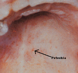
Most cases of infectious mononucleosis are mild or moderate. Supportive therapy including bed rest and analgesics is the primary form of treatment for acute infectious mononucleosis. Symptoms of infectious mononucleosis are very similar to bacterial pharyngitis (caused by Streptococcus pyogenes, which causes strep throat). If a physician treats a patient who he/she thinks has strep throat based solely on clinical signs and symptoms, the physician may give antibiotic treatment to a patient who has infectious mononucleosis. Patients with infectious mononucleosis who are prescribed ampicillin or amoxicillin can develop a rash and mislead the physician into thinking the person is allergic to that class of antibiotic (i.e., beta lactam antibiotics).
The best means of preventing most complications of infectious mononucleosis is exposure to the virus early in life, since the syndrome in children is milder than the disease in adults.
Send comments and email to Dr. Neal R.Chamberlain, nchamberlain@atsu.edu
Revised 11/6/17
©2014 Neal R. Chamberlain, Ph.D., All rights reserved.