Humans have a high level of innate immunity to fungi and most of the infections they cause are mild and self-limiting.
This resistance is due to:
A. Superficial mycoses- infections limited to the outermost layers
of the skin and hair.
| Disease | Etiological Agent | Symptoms | Identification of Organism |
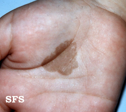
| Pityriasis versicolor | Malassezia furfur | hypopigmented macules | Spaghetti and meatballs appearance of organims in skin scrapings. |
| Tinea nigra | Exophiala werneckii | black macules | Black, 2-celled oval yeast in skin scrapings |
| Black piedra | Piedraia hortai | black nodule on hair shaft | black nodule on hair shaft composed of spore sacs and spores |
| White piedra | Trichosporon beigelii | creme-colored nodules on hair shaft | white nodule on hair shaft composed of mycelia that fragment into arthrospores |
These diseases are restricted to the keritinized layers of the skin,
hair, and nails. Unlike the superficial mycoses, host immune responses
may be evoked, resulting in pathologic changes expressed in the deeper
layers of the skin. The organisms that cause these diseases are called
dermatophytes. These diseases are often called ringworm or tinea. All the
following diseases are causes by
Microsporum,
Trichophyton,
and
Epidermophyton,
which
comprise 41 species.
| Disease | Symptoms | Identification of organism |
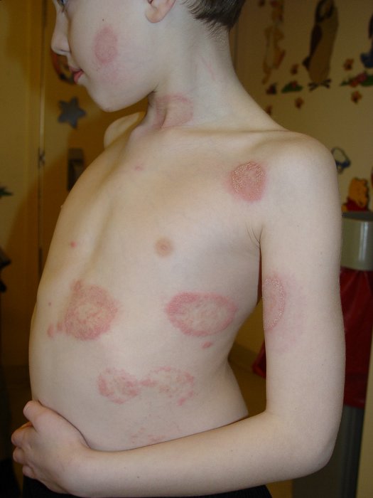
| Tinea capitis | ringworm of scalp | presence/absence and shape of micro- and macroconidia in scrapings of lesion, KOH mount |
| Tinea corporis | ringworm of trunk, arms, legs |
|
| Tinea manuum | ringworm of hand |
|
| Tinea cruris | ringworm of groin "jock itch" |
|
| Tinea pedis | ringworm of foot "athlete's foot |
|
| Tinea unguium | infection of nails |
|
| Ectothrix | infection of hair shaft surface | mycelium and spores on hair shaft |
| Endothrix | infection of hair shaft interior | mycelium and spores in hair shaft |
| Disease | Etiological agent | Symptoms | ID of organism |
| Sporotrichosis | Sporothrix schenckii | Nodules and ulcers along lymphatics and at site of inoculation | Yeast in tissue; mold at rm temp with "rosette pattern" |
| Chromoblastomycosis | Fonsecaea pedrosoi or compacta, Wangiella dermatitidis | warty nodules that progress to "cauliflower-like" appearance a inoculation site. | copper-colored spherical yeasts called "Medlar bodies" in tissue |
| Mycetoma | Pseudallescheria boydii, Madurella grisea or mycetomatis | draining sinus tracts at site of inoculation | white, brown, yellow or black granules in exudate that are fungal colonies |
Histoplasma capsulatum- Ohio and Mississippi river valleys, Yeast cells in tissue, Tuberculate macroconidia in mycelial phase.
Blastomyces dermatitidis- Ohio and Mississippi river valleys, Broad Base Budding yeast in tissue, Mycelia= microconidia
Coccidioides immitis- Southwestern US. Spherule in tissue, barrel-shaped Arthroconidia in mycelia phase.
Cryptococcus neoformans- Only yeast phase but unusual in that the cells are encapsulated as demonstrated by an India Ink stain.
E. Opportunistic mycoses- infections of patients with immune deficiencies who would otherwise not be infected. Ex. AIDS, altered normal flora, diabetes mellitus, immunosuppressive therapy, malignancy.
Candidiasis- Candida albicans- Creamy growth on various body surfaces. ex. mouth, skin, vagina. Budding yeast. Form pseudohyphae in tissue. Germ tube when grown in serum.
Aspergillosis- Aspergillus niger.
TO GO BACK TO MY ![]()
_invasive_type.jpg)