General Goal:To know the major causes of this disease, how it is
transmitted, and understand the basic processes that
result in the progression from HIV infection to AIDS.
Specific Educational Objectives: The student should be able to:
recite the most likely causes of HIV/AIDS and how this viral infection is
usually acquired in the United States (modes of transmission for females and
males are different).
describe how the virus attaches to human cells. Also know the human cell
receptors that the virus attaches to (hint: M-tropic vs. T-tropic viruses).
describe the three different HIV/AIDS disease stages and what happens to the
immune system during those disease stages.
describe the
various means of diagnosing HIV/AIDS and when to use which test. You should also
know CDC's definition for AIDS.
list the most common opportunistic
infections that occur in HIV/AIDS patients.
describe the
basic treatment regimen (HAART).
list ways of preventing
HIV infections (hint: how do you prevent congenital infections?)
Lecture:
Dr. Neal R. Chamberlain
References:
A wonderful and informative website to visit is from
Johns Hopkins Medical Center Called
"The
Body". Go there you will learn alot!
The organisms, or viruses, discussed in this handout can infect several different cell types, including neurons, epithelial cells, salivary glands, lymphocytes, macrophages, and monocytes. The ability of these viruses to infect lymphocytes is discussed according to the dramatic effects the viruses have on the immune response.
There are two basic types of human lymphocytes: B lymphocytes and T lymphocytes. When activated, B lymphocytes become plasma cells that produce antibodies. B lymphocytes require two different signals to become plasma cells: They must bind antigen with their surface bound antibody, and they must be exposed to cytokines produced by T lymphocytes (T-helper cells). Antibody production to foreign antigens is called the humoral immune response.
There are two basic types of T lymphocytes: T-helper cells (CD4+ cells) and T-cytotoxic cells (CD8+ cells). When activated, the T-cytotoxic cells can eliminate the host cells that are infected with virus, Mycobacteria, certain intracellular bacteria, fungi, parasites, and tumor cells. The cellular immune response occurs when cells kill other cells. The T-cytotoxic cells require two signals to become activated. They must bind antigen with their T-cell receptor, and they must be exposed to cytokine (interferon g)from T-helper cells.
There are two types of T-helper cells: Th1 and Th2. These cells are essential in helping the body mount a humoral and a cellular immune response. Th1 T-helper cells produce interferon gamma to activate T-cytotoxic cells. Th2 T-helper cells produce IL-4 and IL-5 to activate B lymphocytes. Without T-helper cells, the patient’s adaptive immune systems (i.e., humoral and cellular immune response) become much less effective at eliminating microbial invaders.
The diseases discussed in this section of the handout are acquired immunodeficiency syndrome (AIDS), infectious mononucleosis, and cytomegalovirus (CMV) infections. Human immunodeficiency virus (HIV) causes AIDS, and the HIV virus infects T-helper lymphocytes (CD4+). CMV causes several different diseases depending on the host infected. CMV can infect many different cell types (e.g., epithelial cells, T cells, macrophages). The infection is spread through the body and establishes a latent infection in T lymphocytes and macrophages. EBV causes infectious mononucleosis and it infects B lymphocytes.
ACQUIRED IMMUNODEFICIENCY SYNDROME
AIDS is an epidemic that has caused significant morbidity and mortality in most countries worldwide. Destruction of CD4+ T lymphocytes (T-helper lymphocytes) predisposes infected individuals to a wide range of opportunistic infections, tumors, dementia, and death.
Etiology
There are two types of HIV virus: HIV type 1 (HIV-1; group M, N, O, and P) and HIV type 2. They are human retroviruses in the lentivirus subfamily. The most common cause of AIDS is HIV-1.
The HIV-1 type contains 4 groups M, N, O, and P. Within the group M there are nine genetically distinct subtypes (clades): A, B, C, D, F, G, H, J, and K (the letters E and I are missing because they were originally thought to be new subtypes but upon further examination they were really CRF viruses; E is now called CRF A/E and I is now called CRF A/G/H/K). Occasionally, two or more subtype viruses of HIV-1 group M can infect the same cell and create a hybrid virus know as a “circulating recombinant form or CRF” (e.g., CRF A/B is a recombinant virus of subtypes A and B).
A new strain of HIV-1 has been reported in patients in Cuba that rapidly progresses to AIDS. After infection with this HIV-1 CRF 19_cpx strain patients progress to AIDS in about 3 years without treatment. Other HIV-1 strains progress to AIDS in about 10 years without treatment. (Vivian et al. 2015. CRF19_cpx is an Evolutionary fit HIV-1 Variant Strongly Associated With Rapid Progression to AIDS in Cuba. Kouri, EBioMedicine, Volume 2, Issue 3, 244-254)
Manifestations
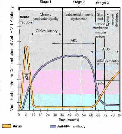
Untreated HIV infection involves three stages of disease (Figure below) and is ultimately fatal in most patients. In many patients, symptoms of HIV infection do not occur until stage 3 of the infection.
Stage 1, or primary HIV infection, has an incubation period of 1–3 weeks before symptoms begin (Table L-1). Stage 1 ends with the production of high titers of anti-HIV antibodies at 2–3 months postinfection.
Table L-1. Symptoms of Stage 1 (Primary HIV Infection)
Patient may have NO symptoms OR they may have any or all of the following:
Encephalitis rash (small pink papules or macules over much of the body)
Stage 2 HIV infection is usually asymptomatic and in most cases lasts for 6 years or longer. Patients produce large amounts of anti-HIV antibody. HIV is detectable in blood, semen, and cervical secretions. If symptoms occur, the patient presents with persistent generalized lymphadenopathy or AIDS-related complex (ARC) (Table L-2).
Table L-2. Symptoms of AIDS-related Complex (ARC)
Fever
Fatigue
Diarrhea
Weight loss
Night sweats
Immunologic abnormalities
Dementia
Spontaneous neoplasms
Stage 3 is usually a period when symptoms of various opportunistic infections or spontaneous neoplasms begin. The severity and frequency of these infections and neoplasms is directly related to the decline of CD4+ T cells.
Course of Infection
Several groups of individuals have been identified based on clinical progression from HIV infection to AIDS. There are rapid progressors, typical progressors, long-term nonprogressors, and highly-exposed persistently seronegative patients. Around 10% of persons infected with HIV are rapid progressors. Rapid progressors are HIV infected patients that develop the symptoms of AIDS within 2-3 years after infection.
Most HIV infected patients fit in the second group of typical progressors. Typical progressors develop AIDS within approximately 10 years after seroconversion.
Another 10% of HIV-positive patients become long-term nonprogressors. Long-term nonprogressors maintain low plasma HIV RNA levels (less than 50 copies/ml) and normal CD4+ T cell counts after more than a decade of HIV seropositivity without drug treatment.
A fourth category is composed of individuals who are multiply-exposed to HIV but do not produce antibodies to HIV and do not have any detectable levels of HIV RNA in their plasma. When these individuals’ peripheral blood monocytes are stimulated with HIV-1 peptides they have lymphoproliferative activity and mount a HIV-1 specific CD8+ CTL activity. This suggests that transient infections appear to have occurred in these persons.
The HIV-1 group M subtype a person is infected with can have an effect on progression of the infection. Persons infected with subtypes C, D, or G develop AIDS sooner than those infected with subtype A if they do not obtain antiretroviral treatment. People infected with CRF A/E progress faster to AIDS than those with subtype B if they don’t get antiretroviral treatment.
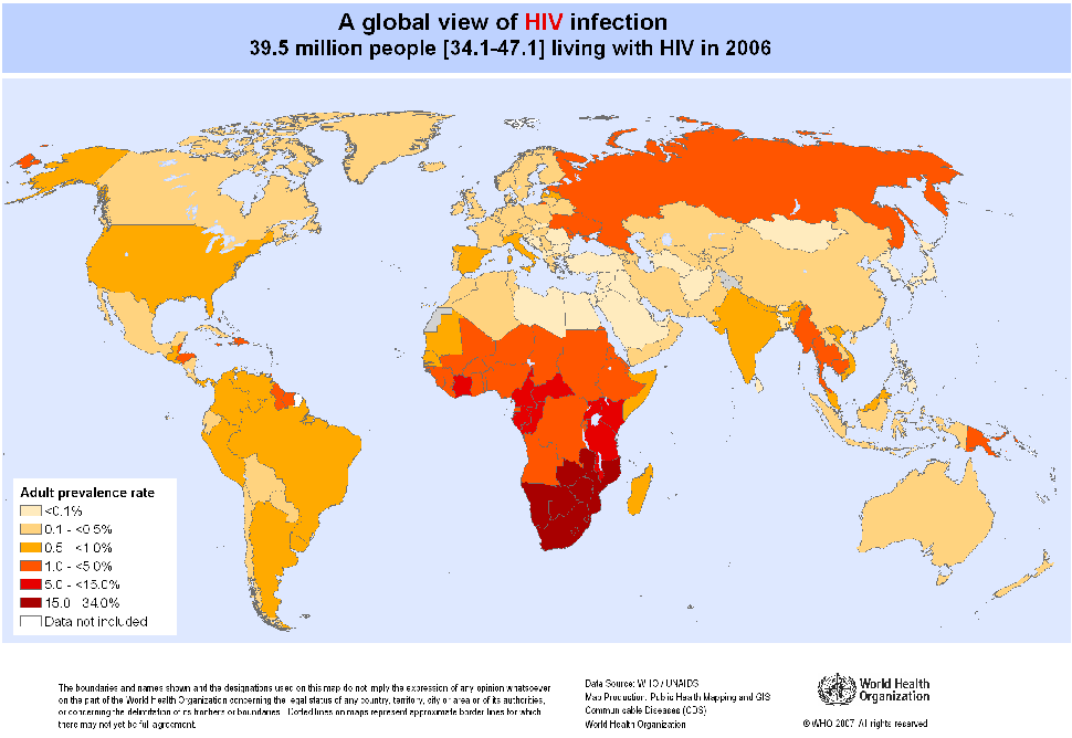
Epidemiology
HIV-1 infection is found worldwide, and HIV-2 is mainly found in West Africa. .
Click on image for an enlarged view.
There are four principal genetic groups of HIV-1, designated M, N, O and P. Genetic group M is highly prevalent and is further classified into 9 subtypes, A, B, C, D, F, G, H, J, and K and CRF recombinant hybrids.
HIV-1 group M subtype B is found predominately in Europe, North and South America, Japan and Australia. HIV-1 group M subtype C is common seen in Southern and East Africa, India, and Nepal. It is responsible for around 50% of the HIV infections in the world.
HIV-1 group M subtype B is spread mostly by homosexual contact and intravenous drug use and HIV-1 group M subtype C is spread by heterosexual contact (i.e., via a mucosal route)
HIV-positive persons are infectious during both asymptomatic and symptomatic stages of infection.
The most common mode of transmission of HIV worldwide is heterosexual contact (Table L-5).
The most common mode of transmission of HIV in males in the US is male-to-male sexual contact, whereas the most common mode of transmission in females is heterosexual contact.
Over 1.1 million persons are living with HIV infection and 1 in 6 do not know they are infected with HIV.
The overall transmission rate is 5 per 100 person years. Transmission rate in undiagnosed persons is 3.5 times higher than in those that know their HIV status.
HIV is NOT transmitted by casual contact or by touching, hugging, kissing, coughing, sneezing, insect bites, water, food, utensils, toilets, and swimming pools or public baths.
Around 50,000 new HIV infection occur each year. Most (636%) of these new infections occurred in gay and bisexual men.
Every 11 minutes and 48 seconds someone is infected with HIV
Homosexual and bisexual men of all races and ethnicities and African American men and women are the groups most affected by HIV.
African Americans, while comprising 13% of the U.S. population, accounted for 44% of the new HIV infections in 2010.
HIV incidence has been steadily increasing among homosexual and bisexual men since the early 1990s.
New HIV infections by heterosexual contact and by injection drug use have been decreasing over time.
HIV disease remains a leading cause of death among persons 25 to 44 years old, particularly among those who are black/African America or Hispanic.
After rapidly increasing since the 1980s, the annual rate of death due to HIV disease peaked in 1994 or 1995, decreased rapidly through 1997, and became level after 1998.
Table L-5. Modes of Transmission of Human Immunodeficiency Virus (HIV)
Transmission
Mode of Transmission
Comments
Sexual
Anal
Vaginal
Homosexual
Heterosexual
Homosexual routes of transmission occur in male homosexuals and are the most common route of transmission in the U.S.
Heterosexual routes of transmission are themost common routes worldwide. Heterosexual routes are also the most common routes of infection in females in the U.S.
Inoculation in blood
Transfusion of blood or blood products
Needle sharing among intravenous drug users
Needlestick, open wound, and mucous-membrane exposure to health care workers (e.g., dentists, oral surgeons)
Tattoo needles
Needle sharing in intravenous drug users is the third most common route of transmission in the U.S.
Perinatal
Transplacental (intrauterine transmission)
Peripartum transmission (during labor and delivery)
Breast milk ingestion in neonates
Peripartum transmission and ingestion of breast milk together are the most common means of transmission to children < 5 years of age
Pathogenesis
The ability of HIV to infect and destroy CD4-expressing T cells (T-helper cells or T-helper lymphocytes) and macrophages induces immunosuppression in patients with AIDS. When large numbers of T-helper cells are destroyed, the body eventually is unable to mount an immune response to infectious agents and to eliminate tumor cells. The severity of the HIV infection is closely aligned with the reduction in CD4+ T cells (T-helper cells) and the increase in HIV virus particles in the blood.
During anal and vaginal intercourse, HIV can bind to both Langerhans and dendritic cells in the epithelium. HIV binds to dendritic cells via a lectin called DC-SIGN (CD209 or C-type lectin receptor). The Langerhans and dendritic cells then transport HIV to the regional lymph nodes or Peyer’s patches and infect the CD4+ T cells. The likelihood of HIV infection being transmitted during anal or vaginal intercourse is higher if the person exposed to an HIV-contaminated secretion already has a sexually transmitted infection.
HIV binds with glycoprotein 120 (gp120) to CD4 T cells in the lymph nodes and uses gp41 to enter the host cells. To infect a CD4+ T cell, gp120 must bind to two host cell surface receptors. All HIV viruses must bind to the CD4 host cell receptor to infect the host cells. However, depending on the strain of HIV virus (i.e., X4 and R5 strains), one of two other host CD4+ T-cell chemokine receptors, known as CCR5 and CXCR4, must be present on the cell to be infected by the virus.
T cell tropic HIV (T-tropic HIV; HIV-1 X4 virus) requires CD4 and CXCR4 host-cell receptors to infect the host cell. T-tropic viruses are usually transmitted via blood and blood products, and are syncytia-inducing viruses that infect CD4+ T cells.
Mucosal surface tropic HIV (M-tropic HIV; HIV-1 R5 virus) requires CD4 and CCR5 host-cell receptors to infect the host cell. M-tropic viruses are usually transmitted via sexual contact. They infect macrophages and some CD4+ T cells, but are not syncytia-inducing viruses.
If HIV is transmitted via percutaneous injection, it can infect dendritic and monocyte-macrophage lineage cells. The macrophage lineage cells produce CD4, CCR5, and CXCR4, which can be infected by M-tropic and T-tropic HIV viruses. The HIV-infected macrophages and dendritic cells can then transport HIV to the regional lymph nodes through the lymph or the bloodstream. Once in the lymph nodes, HIV infects CD4+ T cells.
When HIV reaches the lymph node or Peyer’s patch, it continuously replicates in CD4+ T cells. The virus and infected CD4 T cells are released from the nodes into the blood and then are transmitted to other areas of the body (e.g., lymph nodes, brain, and spleen). HIV can destroy CD4+ T cells in several different ways, including accumulation of the nonintegrated DNA copies of the viral genome, increased permeability of the plasma membrane, syncytia formation, and induction of apoptosis. The host can produce large numbers of CD4+ T cells to replace the cells that are destroyed by HIV. However, without treatment, within 6–10 years the ability of most hosts to replace these cells slows and the number of CD4+ T cells decreases.
CD8+ T cells are critical in controlling the progression of HIV disease. However, to become activated and kill HIV-infected cells or release factors that suppress viral replication, CD8+ T cells must be activated by CD4+ T cells. As the number of CD4+ T cell decreases, so does the number of activated CD8+ T cells. Virus replication is no longer inhibited, and infected cells are not eliminated. The amount of virus in the blood increases, reaching 5000–10,000 viral particles per milliliter of blood.
As the number of CD4+ T cells decreases, the ability of the patient to fight certain infections and eliminate malignant cells also is reduced. CD4+ T cells are essential in activation of CD8+ T cells. CD8+ T cells are important in delayed-type hypersensitivity (DTH) responses, which eliminate viral, fungal, and mycobacterial infections as well as malignant cells. CD4+ T cells also regulate antibody production by B cells. The ability to produce antibodies in response to an infection is reduced, making bacterial infections more common. As the number of CD4+ T cells decreases, HIV-infected monocytes and microglial cells in the brain die and release neurotoxic substances or chemotactic factors that promote inflammation in the brain.
Reservoirs of HIV infection are established early in macrophages and resting T cells during mucosal infection. A pool of latently infected CD4+ T cells develops during the very earliest stages of acute HIV infection. Infected cells are able to persist in the patient’s body for extremely long periods of time, possibly decades.
gp120: HIV recognizes and binds to the
CD4 molecule via viral envelope glycoprotein gp120, and then binds to CXCR4
or CCR-5. A "schematic
drawing" of gp120 binding to the receptors. Diagram of virus "life
cycle".
Diagnosis
There are no unique signs and symptoms of HIV infection, which makes diagnosis difficult unless laboratory tests are, performed (Table L-6). Each year 16-22 million people in the US are tested for HIV infection. The CDC recommends that health care providers test everyone between the ages of 13 and 64 at least once as part of routine health care. Sixteen percent of people in the US who have HIV do not know they are infected. HIV antibodies are usually detectable with an ELISA or other immunoassay (IA; see below) within 3–4 weeks after infection. However, false positives occur, and a second ELISA or IA should be performed; if both tests are positive, a Western blot test or an indirect immunofluorescent assay (IDF) for HIV is necessary to confirm the diagnosis of HIV infection.
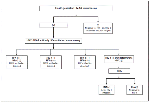
A new fourth generation IA is now available that can detect HIV antibodies (IgM and IgG) and an HIV protein (p24) in the patient’s bloodstream in less than one hour. This test can detect HIV infected patients before their immune system makes antibodies to HIV (early acute HIV infection). It can detect up to 80% of patients in early acute HIV infection. If positive, a Western blot test for HIV is currently necessary to confirm a diagnosis of HIV infection. If the Western blot is positive the patient has antibodies to HIV and is HIV infected. If the Western blot is negative the fourth generation immunoassay may have detected the p24 HIV antigen and an RT-PCR test should be performed to confirm a diagnosis of HIV infection.
The current laboratory diagnostic algorithm for HIV (2 positive ELISA’s then a positive Western blot) cannot detect acute infections and misclassifies approximately 60% of HIV-2 infections as HIV-1. This is because the confirmatory (also called supplemental test) Western blot is less sensitive than the fourth generation IA. A patient during early acute HIV infection will have a positive fourth generation IA however, there is not enough HIV reactive antibodies in the patient’s serum to give a positive Western blot. The Western blot tests for HIV1 and HIV2 will also incorrectly identify HIV2 infected patients as being HIV1 infected.
Therefore, the CDC in June 2013 proposed another way to diagnose HIV infections using two different IA’s; fourth generation IA and a multispot HIV-1/HIV-2 rapid test to determine if the person has HIV-1 or HIV-2 infection. The fourth generation IA can be performed in less than an hour and the multispot HIV-1/HIV-2 rapid test can be performed in about 15 minutes. In many patients this makes serological diagnosis possible during the same day as the patient’s visit. See algorithm below. If the multispot HIV-1/HIV-2 rapid test is negative for both HIV-1 and HIV-2 or is indeterminate then a nucleic amplification test to detect HIV RNA should be performed (RT-PCR).
Peters, PJ, et al. Detection of Acute HIV Infection in Two Evaluations of a New HIV Diagnostic Testing Algorithm — United States, 2011–2013, June 21, 2013, Morbidity and Mortality Weekly Report, 62(24);489-494.
Table L-6. Diagnostic Tests Used to Detect HIV Infection
Test
Purpose
ELISA
Initial screening; two different ELISA results must be positive before a confirmatory test is performed
Latex agglutination
Initial screening
Fourth generation HIV immunoassay
Initial screening; detects HIV antigen (p24) AND antibody to HIV. Can detect patients earlier in the disease process than the ELISA, EIA or latex agglutination tests because it also looks for HIV antigen. Can detect up to 80% of early acute HIV patients (patients that have yet to produce antibody in their bloodstream to HIV)
HIV1/HIV2 differentiation immunoassay
HIV1/HIV2 antibody differentiation test
Western blot analysis
Confirmatory test
p24 antigen
Early marker of infection (detection of a recent infection)
RT-PCR
Detection of virus RNA in blood (detection of a recent infection) and to confirm treatment efficacy
CD4:CD8 T-cell ratio
Staging the disease and to confirm treatment efficacy
Detection of HIV in the blood using reverse transcriptase polymerase chain reaction (RT-PCR) is also considered a confirmatory test for HIV infection. RT-PCR can be used to detect HIV RNA in plasma during the first 2–4 weeks of infection when patients may be seronegative.
To determine if a neonate born to a HIV-infected mother is infected with HIV, an ELISA to detect HIV protein p24 is performed. Antibodies from an HIV-infected mother cross the placenta, making diagnosis of neonatal infections using serology impossible. RT-PCR of neonatal plasma can also be used to detect HIV infection in neonates.
There are now many rapid HIV assays that can be performed with saliva, urine, blood from finger sticks or with blood from venipuncture. These tests can give results in minutes.
HIV-infected patients do not receive a diagnosis of AIDS until they have met the clinical definition of AIDS. The clinical definition for AIDS was developed in 1993 and is useful in treatment decisions and in determining the prognosis of the patient. Tables L-7 through L-10 contain information needed to determine if an HIV-infected patient has AIDS.
Table L-7. The 1993 Revised Classification System for the Diagnosis of HIV Infection and AIDS*
CD4 T-Cell Count
CLINICAL CATEGORIES
(A)
Asymptomatic, Acute (primary) HIV or PGL**
(B)
Symptomatic, Neither Category A nor C Conditions†
(C)
AIDS - indicator Conditions ††
> 500/µL
A1
B1
C1
200–499/µL
A2
B2
C2
< 200/µL
A3
B3
C3
HIV, human immunodeficiency virus; AIDS, acquired immunodeficiency disease.
* All patients who can be classified in the shaded cells of the table have AIDS. Persons with AIDS-indicator conditions (category C; see Table L-10) as well as those with CD4+ T-lymphocyte counts < 200/uL (categories A3 or B3) were reportable as AIDS cases in the United States and territories effective January 1, 1993.
** PGL, persistent generalized lymphadenopathy. See Table L-8 for clinical category A conditions.
† See Table L-9 for clinical category B conditions.
†† See Table L-10 for AIDS indicator conditions.
Table L-8. Clinical Category A Conditions for Diagnosis of HIV Infections*
Consists of one or more of the conditions listed below in an adolescent or adult with documented HIV infection (e.g., two positive ELISA results for HIV and a positive Western blot)
Acute primary HIV infection
Asymptomatic HIV infection
Persistent generalized lymphadenopathy
Accompanying illness or history of acute HIV infection
*Conditions listed in clinical categories B (see Table L-9) and C (see Table L-10) must NOT have occurred.
Table L-9. Clinical Category B Conditions for Diagnosis of HIV Infections*
Symptomatic conditions in an HIV-infected adolescent or adult and that are not included among conditions listed in clinical category C and that meet at least 1 of the following criteria:
The conditions are attributed to HIV infection or are indicative of a defect in cell-mediated immunity, or
The conditions are considered by physicians to have a clinical course or to require management that is complicated by HIV infections.
Category B conditions take precedence over those in category A. For example, someone previously treated for oral or persistent vaginal candidiasis (and who has not developed a category C disease), but who is symptomatic, should be classified in clinical category B.
Examples of conditions in clinical category B include but are not limited to:
Bacillary angiomatosis
Candidiasis, oropharyngeal (thrush)
Candidiasis, vulvovaginal; persistent, frequent or poorly responsive to therapy
Cervical dysplasia (moderate or severe)
Cervical carcinoma in situ
Constitutional symptoms, such as fever (38.5°C) or diarrhea lasting > 1 month
Hairy leukoplakia, oral
Herpes zoster (shingles), involving at least 2 distinct episodes or > 1 dermatome
Idiopathic thrombocytopenia purpura
Listeriosis
Pelvic inflammatory disease, particularly if complicated by tubo-ovarian abscess
Peripheral neuropathy
*Conditions listed in category C (see Table L-10) must not have occurred.
Table L-10. Clinical Category C Conditions for Diagnosis of HIV Infections*
Includes the clinical conditions listed in the AIDS surveillance case definition. For classification purposes, once a category C condition has occurred, the person will remain in category C and is considered to be a patient with AIDS.
Category C conditions include:
Candidiasis of the trachea, bronchi, or lungs
Candidiasis of the esophagus
Cervical carcinoma, invasive
Coccidioidomycosis, disseminated or extra pulmonary
*A patient with any one of these conditions is defined as an AIDS patient regardless of CD4 T-cell count.
Therapy and Prevention
Treatment of an HIV infected patient is complex and requires a strong lifelong commitment from the patient.Multiple studies have shown that better outcomes are achieved in HIV-infected outpatients cared for by a clinician with HIV expertise. Appropriate training, experience, and continuing medical education are essential for optimal patient care. Primary care providers without HIV experience, such as those in rural or underserved areas, should identify experts in the region who can provide consultation when needed.
The following steps are recommended in caring for an HIV infected patient (Panel on Antiretroviral Guidelines for Adults and Adolescents. Guidelines for the use of antiretroviral agents in HIV-1-infected adults and adolescents. Department of Health and Human Services. December 1, 2009; 1-161. Last accessed 9/6/10. http://www.aidsinfo.nih.gov/ContentFiles/AdultandAdolescentGL.pdf):
a. a baseline evaluation
b. laboratory testing for initial assessment and monitoring while on HAART
c. deciding when to start HAART therapy
d. selection of HAART therapy
e. education about HIV risk behaviors and how to prevent HIV transmission to others.
A. Baseline Evaluation
Each HIV-infected patient should have a complete medical history, physical examination, laboratory evaluation, and counseling regarding the implications of HIV infection. The evaluation must include assessment of substance abuse, economic factors (e.g., unstable housing), social support, mental illness, comorbidities, high-risk behaviors, and other factors that are known to impair the ability to adhere to treatment and to promote HIV transmission.
Laboratory tests:
HIV antibody testing (if prior documentation is not available or if HIV RNA is undetectable);
CD4+ T-cell count;
Plasma HIV RNA (viral load);
Complete blood count, chemistry profile, transaminase levels, BUN and creatinine, urinalysis, screening test for syphilis (e.g., RPR, VDRL, or treponema EIA), tuberculin skin test (TST) or interferon-γ release assay (IGRA) (unless there is a history of prior tuberculosis or positive TST or IGRA), anti-Toxoplasma gondii IgG, hepatitis A, B, and C serologies, and Pap smear in women;
Fasting blood glucose and serum lipids if the patient is considered at risk of cardiovascular disease and for baseline evaluation prior to initiation of combination antiretroviral therapy; and
For patients who have pretreatment HIV RNA >1,000 copies/mL, genotypic resistance testing when the patient enters into care, regardless of whether therapy will be initiated immediately. For patients who have HIV RNA levels of 500–1,000 copies/mL, resistance testing also may be considered, even though amplification may not always be successful. If therapy is deferred, repeat testing at the time of antiretroviral initiation.
Testing for Chlamydia trachomatis and Neisseria gonorrhoeae is encouraged to identify both recent high-risk sexual behavior and the need for sexually transmitted disease (STD) therapy;
Chest x-ray in the presence of pulmonary symptoms or with a positive TST or IGRA test.
B.Laboratory testing for initial assessment and monitoring while on HAART
Two surrogate markers are used routinely to assess the immune function and level of HIV viremia: CD4+ T-cell count and plasma HIV RNA (viral load). These tests should be given at initiation of HAART therapy and every 3-6 months during HAART therapy. The CD4+ T-cell count is a major clinical indicator of immune function in patients who have HIV infection. It is one of the key factors in deciding whether to initiate antiretroviral therapy and chemoprophylaxis for opportunistic infections, and is the strongest predictor of subsequent disease progression and survival according to clinical trials and cohort studies. Plasma HIV RNA (viral load) should be measured in all patients at baseline and on a regular basis in patients who are on HAART because viral load is the most important indicator of response to HAART. Plasma viremia is a strong prognostic indicator in HIV infection. Real time PCR is used to determine HIV-1 RNA levels in the blood.
A CBC with differential should be obtained every 3-6 months. Blood chemistry, AST, ALT, and bilirubin should be obtained every 6-12 months.
C. Deciding when to start HAART therapy
HAART should be offered toall HIV-infected individuals. The strength of the recommendation varies on the basis of the pretreatment CD4+ cell count. The LOWER the CD4 cell count the STRONGER the recommendation to begin HAART therapy.
Resistance testing should be used to guide selection of an antiretroviral regimen in both treatment-naïve and treatment-experienced patients; a viral tropism assay should be performed prior to initiation of a CCR5 antagonist; and HLA-B*5701 testing should be obtained prior to initiation of abacavir (they are hypersensitive to this drug).
D. Selection of HAART therapy
Eradication of HIV infection cannot be achieved with currently available antiretroviral regimens; therefore, lifelong treatment to suppress the virus is necessary. Highly active antiretroviral therapy (HAART), available since 1995, has resulted in durable antiviral responses and many benefits of long-term therapy are being reported. Successful HAART results in suppression of viral replication and halts damage to the immune system. It also partially restores the immune system, leading to partial restoration of immune function. Clinical benefits accompanying these immunologic benefits include fewer opportunistic infections and a longer lifespan for patients. Six different classes of antiretroviral drugs are available and are listed in Table L-11.
Table L-11. Antiretroviral Drugs Used in HAART*
Class of Antiretroviral Drug
Mechanism of Action
Drug Names
Nucleoside or nucleotide reverse transcriptase inhibitors (NRTIs)
NRTIs inhibit HIV’s reverse transcriptase and can be placed within the viral DNA. When the NRTIs are placed in the viral DNA by the reverse transcriptase, transcription of the viral genes is inhibited. This prevents virus replication and subsequent spread of the viral infection.
These drugs also inhibit reverse transcriptase, which prevents virus multiplication and spread.
Efavirenz (EFV), etravirine, nevirapine, delavirdine, and rilpivirine
Protease inhibitors (PIs)
HIV produces its own protease, which is important in the production of infective viral particles. The protease cleaves the viral proteins to the correct sizes so that a mature viral particle can form (viral assembly). The PIs inhibit the retroviral protease from cleaving the viral proteins. These drugs help to slow the spread of the virus to other uninfected cells.
A peptide that interferes with the viral gp41 protein and prevents fusion of HIV with the host cell.
Enfuvirtide
CCR5 entry inhibitors
These inhibitors bind to the CCR5 receptor on the host CD4 cells and block binding of the HIV virion to the surface of the CD4 cells.
Maraviroc
Integrase inhibitor
This drug inhibits the enzyme that integrates HIV genetic material into the host chromosome.
Raltegravir; raltegravir 400 mg twice daily plus emtricitabine plus tenofovir suggested for treatment naïve HIV patients
(elvitegravir and dolutegravir- Phase III trials; 2010)
*HAART, highly active antiretroviral therapy. Note: This list is likely to be incomplete because new antiretroviral drugs are rapidly being approved.
Design a treatment regimen for treatment of naïve patients: NNRTI OR a PI OR an integrase inhibitor PLUS 2 NRTIs. Four regimens are now listed as “Preferred” regimens for treatment-naïve patients. They are:
E. Education about HIV risk behaviors and how to prevent HIV transmission to others. This information should be given during the patient’s initial visit and they should be reminded periodically of risky behaviors and how to prevent transmission to others.
Prevention
1. Screen for HIV infection in ALL persons from 13 to 64 years of age
2.Treat HIV infected persons with HAART
Studies have shown that HAART can reduce transmission by HIV-infected persons (Chou R, Selph S, Dana T, et al. Screening for HIV: systematic review to update the 2005 U.S. Preventive Services Task Force recommendation. Ann Intern Med 2012;157:706-718).
3.PreExposure Prophylaxis (PrEP)- November 2010, NIH announced the results of the iPrEx clinical trial, a large, multi-country research study examining PrEP. This study found that daily oral use of tenofovir plus emtricitabine (TDF/FTC, brand name Truvada) provided an average of 44% additional protection to men who have sex with men (MSM). These men also received a comprehensive package of prevention services that included monthly HIV testing, condom provision, and management of other sexually transmitted infections.
4.On-Demand PrEP- A recent study demonstrated 86% reduction in HIV infections in MSM using on-demand PrEP. This involves the person taking two Truvada (tenofovir/emtricitabine, Gilead Science) tablets 2-24 hours before sex and one tablet 24 hr and 48 hr after sex (Molina JM, et al. Abstract #LB23. On Demand PrEP With Oral TDF-FTC in MSM: Results of the ANRS Ipergay Trial. Presented at: Conference on Retroviruses and Opportunistic Infections; Feb. 23-26, 2015; Seattle.)
July 2011, a CDC study called the TDF2 study, along with a separate trial by the University of Washington, provided evidence that a daily oral dose of antiretroviral drugs used to treat HIV infection can reduce HIV acquisition among uninfected individuals exposed to the virus through heterosexual sex. They also used daily oral dose of tenofovir plus emtricitabine (TDF/FTC, brand name Truvada) and obtained a reduction in HIV infections of 63%.
NOW in high risk HIV negative heterosexual and homosexual individuals you can encourage them to take a daily dose of tenofovir plus emtricitabine (TDF/FTC, brand name Truvada) to reduce their chances of getting HIV. Please also note that you should encourage safe sex practices and manage any other sexually transmitted infections they might acquire.
2010 Prevention study: A tenofovir-based microbicide gel may reduce the incidence of new HIV infection by nearly 40% and cut herpes incidence in half in a cohort of heterosexual women in South Africa, according to results presented at the 18th International AIDS Conference, July 2010. Eligible women were aged 18 to 40 years, sexually active, did not have HIV and lived in KwaZulu-Natal, South Africa. There were 445 women randomly assigned to receive the study drug and 444 women assigned to placebo.
In the double blind trial, CAPRISA 004, women were instructed to apply the gel or placebo up to 12 hours before sexual activity and a second dose as soon as possible after sexual activity (no later than 12 hours after sexual activity) for a maximum of two doses in a 24 hr period. The researchers analyzed results for monthly pregnancy tests, quarterly pelvic exams and blood samples taken at months 3, 12, 24 and study exit at 30 months. During monthly visits, all participants were provided with HIV risk-reduction counseling, condoms and treatment for sexually transmitted infections, and each was clinically examined for potential side effects and tested for HIV infection. Results indicated that HIV incidence was 5.6 per 100 women-years (38 infections in 680.6 women years) in the tenofovir arm and 9.1 per 100 women-years (60 infections in 660.7 women-years) in the placebo arm (incidence rate ratio, 0.61; P=.017).
Among women who reported using the gel 80% of the time or more, HIV incidence was 54% lower in the tenofovir group (P=.025). Also in the tenofovir group, intermediate adherers, defined as women with 50% to 80% adherence rates, were 38% less likely to acquire HIV, and low adherers, defined as women with less than 50% adherence, were 28% less likely to become infected. Overall, tenofovir use reduced incidence of new HIV infection by 39%. The CAPRISA 004 study is only a first step in determining if tenofovir gel is effective in preventing HIV and herpes infection; additional studies are needed to confirm and extend the findings of the CAPRISA study.
There is no vaccine currently available to prevent HIV/AIDS. Prevention methods that have reduced the incidence of HIV infections include safe-sex practices (condom use), safe use of needles (no needle sharing), and early screening for HIV infection. Heterosexual circumcised men are less likely to acquire HIV infections than uncircumcised men. Circumcision reduces female-to-male transmission by about 50%; however, circumcision does not appear to prevent HIV transmission in homosexual males.
Treatment of HIV-1 infected pregnant women, as indicated below, can prevent most infections of the fetus or infant (Table L-13).
Table L-13. Treatment to Prevent Transmission of HIV from an HIV-infected Mother to the Fetus or Infant
Time of Zidovudine(ZDV)Administration
Regimen
Antepartum
Oral administration of ZDV to the mother initiated at 14–34 weeks’ gestation and continued throughout the pregnancy
Intrapartum
Intravenous administration of ZDV to the mother during labor and until delivery and single dose of nevirapine during labor
Postpartum
A single dose of nevirapine to the newborn after birth and oral administration of ZDV to the newborn for the first 6 weeks of life, beginning 8–12 hours after birth